Abstract
Minimally invasive right mini-thoracotomy is conventionally performed with femoro-femoral cardiopulmonary bypass (CPB) cannulation. Central (aorto-caval) cannulation through the thoracotomy window, though technically demanding, avoids the morbidity associated with femoral access and does not require specialised minimally invasive equipments. Its applicability to combined congenital lesions — particularly atrial septal defect (ASD) with pulmonary stenosis (PS) — has not been widely described. Here we discuss the case of a 20-year-old female presenting with ostium secundum ASD and severe PS. She underwent combined intracardiac repair of ASD with a Dacron patch and relief of PS via pulmonary arteriotomy and right ventricular outflow tract (RVOT) resection through a right mini-thoracotomy approach with conventional central (aorto-caval) cannulation, without femoro-femoral bypass. Aortic cross-clamp time was 38 minutes, cardiopulmonary bypass duration 65 minutes, post-operative extubation at 6 hours, and total drain output 100 mL. There were no procedure-related complications. Post-operative echocardiography confirmed no residual ASD shunt and satisfactory relief of right ventricular outflow obstruction. This report emphasizes that combined ASD repair and PS relief via right mini-thoracotomy with central cannulation is safe, reproducible and technically feasible without dedicated minimally invasive equipment. This approach minimises sternotomy-related morbidity, reduces cost and yields excellent cosmetic results — particularly advantageous in young female patients.
Keywords
Mini-thoracotomy, Atrial Septal Defect, Pulmonary Stenosis, Central Cannulation, RVOT,
Minimally Invasive Cardiac Surgery (MICS), Congenital Heart Disease
1. Introduction
Minimally invasive cardiac surgery (MICS) has become increasingly preferred over conventional median sternotomy for a range of congenital and acquired cardiac procedures, offering patients reduced post-operative pain, shorter ICU and hospital stays, lower infection risk, and significantly superior cosmetic outcomes. The right mini-thoracotomy approach, in particular, has been established as a reliable route for ASD closure, mitral and tricuspid valve surgery, and excision of intracardiac tumours.
The prevailing paradigm for right mini-thoracotomy mandates femoro-femoral cardiopulmonary bypass (CPB) cannulation. This approach, while effective, carries inherent disadvantages including risk of femoral vessel injury, retrograde aortic perfusion, limb ischaemia, and the requisite use of specialised equipment — elongated femoral cannulae, double-lumen endotracheal tubes, trans-oesophageal echocardiography (TOE), and video-assisted visualisation — each adding to operative complexity and cost. In resource-limited public hospital settings, these requirements pose significant logistical and financial barriers to offering minimally invasive surgery.
Our group at Sawai Man Singh Medical College, Jaipur has developed and validated a technique of conventional central (aorto-caval) cannulation through the mini-thoracotomy window itself, demonstrated across a spectrum of cardiac pathologies including ventricular septal defect (VSD) repair, valve surgery, and left atrial myxoma excision. The current case extends this experience to a combined congenital lesion — secundum ASD with severe PS — representing, to our knowledge, a technically novel application of this cannulation strategy.
Pulmonary stenosis, when combined with ASD, produces a characteristic haemodynamic milieu of right ventricular pressure overload with eventual right-to-left shunting. Surgical correction requires both ASD closure and relief of RVOT obstruction, typically demanding broad access. This case demonstrates that both objectives can be accomplished through a limited 3-inch sub-mammary incision using standard cardiac surgical instruments, without femoral cannulation or video assistance.
2. Case Presentation
A 20-year-old female was referred to our department with complaints of progressive exertional dyspnoea and palpitations over 18 months. Transthoracic echocardiography revealed a large secundum ASD (defect diameter ~20 mm, Qp:Qs ~1.8:1) with concurrent severe valvar and infundibular pulmonary stenosis (peak RVOT gradient 78 mmHg, peak pulmonary valve gradient 62 mmHg; pulmonary valve area reduced with doming, thickened leaflets). Right ventricular hypertrophy and mild right atrial dilatation were noted. There was no evidence of right-to-left shunting at rest. Pulmonary vascular resistance was within acceptable range.
Clinically, the patient was in New York Heart Association (NYHA) Class II–III. Chest X-ray showed cardiomegaly with a prominent pulmonary artery segment. ECG demonstrated right axis deviation and right ventricular hypertrophy pattern. Pulmonary function tests and routine laboratory investigations were within normal limits.
After multidisciplinary evaluation, combined surgical repair was planned via right mini-thoracotomy with conventional central cannulation, avoiding sternotomy. The patient and family were thoroughly counselled regarding the operative plan, its advantages, and potential risks. Written informed consent was obtained.
2.1. Surgical Technique
2.1.1. Patient Positioning
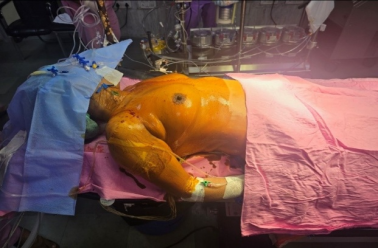
The patient was positioned supine on the operating table. A firm, wedge-shaped pad was placed beneath the right scapula to elevate the right hemithorax 30–45 degrees, improving lateral chest exposure while preserving anaesthetic access (
Figure 1). Standard general anaesthesia was induced using a single-lumen endotracheal tube. Arterial line, central venous catheter, and urinary catheter were placed. No double-lumen tube, trans-oesophageal echocardiography probe, or external defibrillator pads were used.
Figure 1. Patient positioning.
2.1.2. Incision and Thoracic Entry
A sub-mammary antero-lateral skin incision of approximately 3 inches (7–8 cm) was fashioned in the natural inframammary crease, providing excellent cosmetic concealment. The pectoralis major muscle was partially retracted medially without division. The third intercostal space was entered along the superior border of the fourth rib, avoiding injury to the neurovascular bundle. A narrow-blade rib retractor was inserted and gently opened. The right lung was retracted laterally by the anaesthetist through brief single-lung ventilation periods, without the need for a double-lumen tube.
2.1.3. Pericardiotomy and Vascular Exposure
Pericardiotomy was performed 2 cm anterior to and parallel to the right phrenic nerve, with the incision extended superiorly and inferiorly to maximise vascular exposure. This manoeuvre exposed the ascending aorta, the superior vena cava (SVC), and the inferior vena cava (IVC) satisfactorily. The SVC and IVC were individually encircled with umbilical tape to allow snaring during bicaval CPB. The main pulmonary artery was additionally identified and looped in anticipation of pulmonary arteriotomy.
2.1.4. Heparinisation and Cannulation
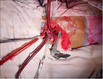
Systemic heparin was administered (target ACT >480 seconds). Purse-string sutures were placed on the ascending aorta (2-0 prolene, felt-pledgetted), SVC, and IVC under direct vision. Particular care was taken during aortic manipulation given the limited thoracotomy window. The ascending aorta was cannulated centrally in standard fashion with an arterial cannula; separate bicaval venous cannulae were inserted into the SVC and IVC (
Figure 2). No femoral cannulae or specialised elongated cannulae were required.
Figure 2. Aortic and bicaval cannulation.
2.1.5. Cardiopulmonary Bypass and Myocardial Protection
Cardiopulmonary bypass was established and the patient cooled to mild hypothermia (32°C). After satisfactory venous drainage, the SVC and IVC were snared. The ascending aorta was cross-clamped and antegrade cold blood cardioplegia was delivered via the aortic root. Satisfactory diastolic cardiac arrest was achieved promptly.
2.1.6. ASD Repair via Right Atriotomy
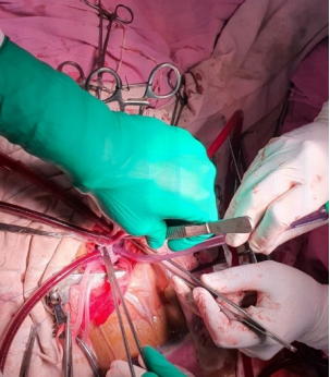
The right atrium was opened via a standard right atriotomy parallel to the atrioventricular groove. Intracardiac anatomy was inspected. A large secundum ASD was confirmed, with well-defined margins and no deficiency of the anterosuperior or posteroinferior rims (
Figure 3). A pre-cut Dacron patch of appropriate size was sutured to close the ASD using continuous 4-0 polypropylene suture, anchored with interrupted pledgeted sutures at the posteroinferior rim adjacent to the atrioventricular node and coronary sinus. The right atriotomy was temporarily partially closed to allow pulmonary artery work.
Figure 3. Atrial septal defect.
2.1.7. Relief of Pulmonary Stenosis via Pulmonary Arteriotomy and RVOT
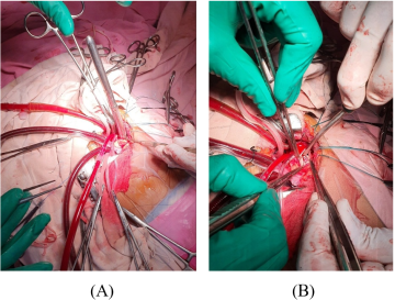
The main pulmonary artery was opened via a longitudinal pulmonary arteriotomy, extended inferiorly across the pulmonary annulus into the RVOT where required. The pulmonary valve leaflets were found to be thickened, fused at the commissures, and domed in systole — consistent with valvar pulmonary stenosis (
Figure 4A). Commissurotomy was performed along the fused commissures with a scalpel, taking care to preserve leaflet mobility. Infundibular fibromuscular bands causing subvalvar obstruction within the RVOT were resected under direct vision through the arteriotomy and the right atriotomy. Hegar dilatation of the pulmonary valve was done to ensure adequacy of the valve (
Figure 4B). The pulmonary arteriotomy was closed with continuous 5-0 polypropylene suture. Satisfactory reduction in annular and infundibular obstruction was confirmed on digital palpation.
Figure 4. (A). Pulmonary Stenosis (B). Hegar dilatation of Pulmonary Stenosis.
The right atriotomy was then fully closed in two layers with 4-0 polypropylene. De-airing manoeuvres were performed via needle aspiration of the ascending aorta and the pulmonary artery. The aortic cross-clamp was released and spontaneous defibrillation to sinus rhythm was achieved.
2.1.8. Weaning, Decannulation and Closure
The patient was weaned from cardiopulmonary bypass without pharmacological inotropic support. Haemodynamics were stable. Decannulation was performed in reverse order. Protamine sulphate was administered to reverse heparinisation. Meticulous haemostasis was achieved. The pericardium was partially re-approximated with absorbable sutures. A single pleural drain was positioned. The thoracotomy was closed in layers — ribs with heavy absorbable pericostal sutures, musculature with continuous vicryl, and skin with subcuticular absorbable suture for the best cosmetic result.
2.2. Operative Data Summary
Table 1. Various parameters pertaining to the patient and intraoperative and postoperative variables, with their values and clinical significance with regards to the patient.
Parameter | Value | Clinical Significance |
Age / Sex | 20 years / Female | Young patient — cosmesis critical |
Diagnosis | Secundum ASD + Severe PS | Combined congenital lesion |
Approach | Right mini-thoracotomy (3rd ICS) | Sub-mammary incision |
Cannulation | Central aorto-caval | No femoral access needed |
ASD repair | Dacron patch closure | Standard patch technique |
PS relief | Pulmonary arteriotomy + RVOT resection | Combined infundibular + valvar relief |
Cross-clamp time | 38 minutes | Comparable to sternotomy series |
CPB duration | 65 minutes | Acceptable bypass time |
Extubation | 6 hours post-op | Fast-track recovery |
Drain output | 100 mL | Minimal blood loss |
Complications | None | Safe uneventful course |
3. Results
The combined procedure was completed without intraoperative complications. Cannulation was smooth and satisfactory intracardiac exposure was maintained throughout both stages — ASD patch closure and pulmonary arteriotomy with RVOT resection — via the limited thoracotomy window.
Total aortic cross-clamp time was 38 minutes and cardiopulmonary bypass duration was 65 minutes. The patient was extubated 6 hours post-operatively. Total drain output over 24 hours was 100 mL. She was transferred to the ward on post-operative day 2 and discharged on post-operative day 6.
Post-operative transthoracic echocardiography confirmed complete closure of the ASD with no residual shunting. The peak RVOT gradient reduced to 18 mmHg, with satisfactory pulmonary valve opening and no significant pulmonary regurgitation. Right ventricular systolic pressure normalised progressively on serial follow-up. At 6-week clinic review, the patient was in NYHA Class I with an excellent cosmetic incision concealed within the inframammary fold.
4. Discussion
Surgical repair of ASD is one of the most common congenital cardiac operations performed worldwide. In isolation, ASD closure via right mini-thoracotomy has been well-described, with multiple institutional series confirming safety and reproducibility. However, the concomitant presence of pulmonary stenosis — requiring pulmonary arteriotomy and RVOT resection in addition to ASD patch closure — introduces a significant increment in operative complexity when approached through a limited incision.
The standard teaching holds that complex combined congenital repairs may necessitate wider access — either sternotomy or, at minimum, peripheral CPB cannulation to optimise visualisation. This case challenges this presumption, demonstrating that both ASD closure and PS relief (combined valvar commissurotomy and infundibular resection) can be accomplished through a 3-inch sub-mammary incision with central cannulation, without any adjunctive minimally invasive technology.
Central aorto-caval cannulation through the mini-thoracotomy window is the cornerstone of our institutional technique
| [1] | Lukram S, Sharma M, Dixit S, Sharma A. Submammary incision as mini right thoracotomy approach with intercostal nerve exclusion technique in adjunction with intercostal nerve blockage: a comparative study in young female in Indian population undergoing mitral valve replacement. Heart India. 2016, 4, 61–66. |
[1]
. As reported across our cumulative experience — spanning VSD repair
| [2] | Dixit S, Sharma A, Suthar J, Watti V, Sharma M. Repair of ventricular septal defect through anterolateral thoracotomy with central cannulation: our experience. Indian Journal of Thoracic and Cardiovascular Surgery. 2020, 36(5), 476–482.
https://doi.org/10.1007/s12055-020-00929-w |
| [3] | Sharma A, Dixit S, Sharma M, Suthar J. Mini-thoracotomy approach with central cannulation for repair of ventricular septal defects — a better alternative. Journal of Cardiothoracic Surgery. 2019, 14, 065. |
[2, 3]
, mitral valve surgery
| [5] | Sharma A, Dixit S, Sharma M, Suthar JK, Mittal S. Anterolateral minithoracotomy mitral valve surgery with central cannulation: a three-year single-center experience. Heart Views. 2024; 25(3): 127–132.
https://doi.org/10.4103/heartviews.heartviews_10_24 |
[5]
, double valve replacement
| [4] | Sharma A, Dixit S, Sharma M, Suthar JK, Mittal S. Right thoracotomy with central cannulation for valve surgery: 10 years of experience. J Cardiothorac Surg. 2024; 19: 597.
https://doi.org/10.1186/s13019-024-02945-y |
| [7] | Sharma A, Dixit S, Mittal S, Sharma M, Sharma D, Mawar KK. Del-Nido cardioplegia versus St Thomas cardioplegia solution in double valve replacement: a single-centre experience. Perfusion. 2021, 36(4).
https://doi.org/10.1177/0267659120961921 |
[4, 7]
, left atrial myxoma excision
| [6] | Pandey GM, Sharma A, Dixit S, Sharma M, Mittal S, Singh P. Minimally invasive approach for excision of left atrial myxoma — an 8-year single-center experience. Indian Journal of Cardiovascular and Thoracic Surgery. 2026, 42(7), 913-917.
https://doi.org/10.1007/s12055-026-02184-x |
[6]
, and now combined ASD+PS — this cannulation strategy is not merely feasible but consistently safe and reproducible across a spectrum of pathologies when performed by a trained team. The key technical requirements are: adequate sub-mammary incision in the 3
rd intercostal space, pericardiotomy anterior to the right phrenic nerve, direct-vision aortic cannulation with felt-pledgetted purse strings, and bicaval cannulation with standard-length venous cannulae.
The pulmonary artery, approached via the mini-thoracotomy window, is accessible through the same exposure used for aortic and caval cannulation. The main pulmonary trunk lies anterior and to the left of the aorta; through the right thoracotomy approach, it can be identified, looped, and opened via a vertical arteriotomy with excellent visualisation. Infundibular RVOT resection, performed through a combination of the arteriotomy incision and the right atriotomy, provides adequate working space without requiring trans-ventriculotomy, thereby avoiding right ventriculotomy-related morbidity.
The sub-mammary approach for young female patients has previously been validated in many series, especially one where sex-differentiated approach was used for male and female patients
| [8] | Vida VL, Padalino MA, Boccuzzo G, Stellin G. Minimally invasive operation for congenital heart disease: a sex-differentiated approach. Journal of Thoracic and Cardiovascular Surgery. 2009, 138(4), 933–936. |
[8]
. Similarly in our group, the sub-mammary approach has demonstrated superiority over standard lateral thoracotomy or minimal sternotomy approach
| [9] | Nicholson IA, Bichell DP, Bacha EA, del Nido PJ. Minimal sternotomy approach for congenital heart operations. Annals of Thoracic Surgery. 2001, 71(2), 469–472. |
[9]
in terms of pain scores, cosmesis
| [10] | Bleiziffer S, Schreiber C, Burgkart R, et al. The influence of right anterolateral thoracotomy in prepubescent female patients on late breast development and the incidence of scoliosis. Journal of Thoracic and Cardiovascular Surgery. 2004, 127(5), 1474–1480. |
| [15] | Sabzi F, Hama Tofiq PA, Asadmobini A, Solouki L. Comparative Study of Surgical Complications in Atrial Septal Defect Repair: Right Anterior Mini-thoracotomy Versus Classic Midline Sternotomy. International Cardiovascular Research Journal. 2025, 19(1): e157987.
https://doi.org/10.5812/icrj-157987 |
[10, 15]
, and patient satisfaction. However one study quoted impaired breast development after anterolateral thoracotomy in a female patient following ASD repair and advocated use of a midaxillary approach in pre-pubescent females
| [11] | Schreiber C, Bleiziffer S, Kostolny M, et al. Minimally invasive midaxillary muscle-sparing thoracotomy for ASD and PAPVC repair in prepubescent female patients. Annals of Thoracic Surgery. 2005, 80(5), 1876–1879. |
[11]
. In our approach, the combined use of intercostal nerve preservation techniques and nerve blockade further reduces post-thoracotomy pain. In the current case, the same incision philosophy was applied to a congenital repair scenario, with equally satisfactory cosmetic and functional outcomes. Apart from cosmesis, short ICU and hospital stay
| [12] | Comparative Study between Anterolateral Thoracotomy and Standard Median Sternotomy for Atrial Septal Defect (ASD) Closure. The Egyptian Journal of Hospital Medicine. 2026, 103(1), 2352-2358.
https://doi.org/10.21608/ejhm.2026.502876 |
[12]
, the risk of complications like arrythmia and atelectasis are also less
| [13] | Ullah A, Zaher A, Saha H, Mamun S, Akhter S. Comparison of Short-Term Outcomes: Minimally Invasive Thoracotomy Versus Median Sternotomy for Atrial Septal Defect Closure. Cureus 17 (1), 2025. https://doi.org/10.7759/cureus.77898 |
[13]
, with zero thirty day mortality in a recent meta-analysis
| [14] | EL-Andari R, Watkins AR, et al. A Systematic Review of Minimally Invasive Approaches to Surgical Atrial Septal Defect Repair. Heart, Lung and Circulation. 2025, Volume 34, Issue 8, 764-776. https://doi.org/10.1016/j.hlc.2025.01.010 |
[14]
.
From a resource perspective, the elimination of femoral cannulae, double-lumen tubes, TOE, and video-assisted instrumentation represents a significant cost reduction. In government-sector tertiary care hospitals serving predominantly rural and lower-income populations — such as Sawai Man Singh (SMS) Medical College, Jaipur — this cost advantage is not merely convenient but critically important for widening access to minimally invasive surgery beyond high-resource centres.
Cross-clamp (38 minutes) and CPB time (65 minutes) times in this case are comparable to, and in some respects shorter than, equivalent combined repairs performed via sternotomy in many published series — underscoring that central cannulation through mini-thoracotomy does not meaningfully prolong operative time once institutional experience is established.
Regarding haemodynamic considerations specific to the ASD+PS combination: the presence of severe PS with a high RVOT gradient (>70 mmHg) raises the theoretical concern of inadequate right ventricular decompression during CPB. In our case, bicaval cannulation with adequate venous drainage and prompt cardioplegic arrest provided satisfactory right heart decompression. The pulmonary artery was vented intraoperatively to prevent right heart distension.
Potential limitations of this approach include its learning curve, the requirement for a skilled and experienced operative team, and the absence of intraoperative TOE for real-time guidance. We address the latter by using post-bypass digital palpation of the pulmonary artery and direct aortic root needle de-airing, with post-operative echocardiography as the definitive assessment tool.
5. Conclusion
Combined surgical repair of atrial septal defect with pulmonary stenosis relief — encompassing Dacron patch ASD closure, pulmonary valve commissurotomy, and RVOT infundibular resection — can be safely and effectively accomplished via a right mini-thoracotomy with conventional central aorto-caval cannulation. This approach avoids sternotomy, femoral vessel access, and dedicated minimally invasive equipment, making it practical and cost-effective in resource-limited settings.
The excellent operative outcome — zero complications, early extubation, minimal blood loss, short ICU and hospital stay, and complete haemodynamic correction — validates this technique as a reproducible strategy for combined congenital repair. It is particularly well-suited to young female patients for whom cosmetic outcome is a priority. Wider dissemination of this technique, supported by the growing body of institutional evidence from our centre, offers the potential to democratise minimally invasive congenital cardiac surgery in the public health sector.
Abbreviations
CPB | Cardio Pulmonary Bypass |
ASD | Atrial Septal Defect |
PS | Pulmonary Stenosis |
RVOT | Right Ventricular Outflow Tract |
MICS | Minimally Invasive Cardiac Surgery |
TOE | Trans Oesophageal Echocardiography |
VSD | Ventricular Septal Defect |
NYHA | New York Heart Association |
SVC | Superior Vena Cava |
IVC | Inferior Vena Cava |
SMS | Sawai Man Singh |
Author Contributions
Gaurav Manglam Pandey: Visualization, Writing – original draft, Writing – review & editing
Anil Sharma: Conceptualization, Data curation, Formal Analysis, Methodology, Supervision, Validation, Writing – review & editing
Sunil Dixit: Methodology, Supervision, Validation, Visualization, Writing – review & editing
Mohit Sharma: Supervision, Validation, Visualization, Writing – review & editing
Sourabh Mittal: Supervision, Validation, Visualization, Writing – review & editing
Pritanshi Singh: Writing – review & editing
Conflicts of Interest
The authors declare no conficts of interest.
References
| [1] |
Lukram S, Sharma M, Dixit S, Sharma A. Submammary incision as mini right thoracotomy approach with intercostal nerve exclusion technique in adjunction with intercostal nerve blockage: a comparative study in young female in Indian population undergoing mitral valve replacement. Heart India. 2016, 4, 61–66.
|
| [2] |
Dixit S, Sharma A, Suthar J, Watti V, Sharma M. Repair of ventricular septal defect through anterolateral thoracotomy with central cannulation: our experience. Indian Journal of Thoracic and Cardiovascular Surgery. 2020, 36(5), 476–482.
https://doi.org/10.1007/s12055-020-00929-w
|
| [3] |
Sharma A, Dixit S, Sharma M, Suthar J. Mini-thoracotomy approach with central cannulation for repair of ventricular septal defects — a better alternative. Journal of Cardiothoracic Surgery. 2019, 14, 065.
|
| [4] |
Sharma A, Dixit S, Sharma M, Suthar JK, Mittal S. Right thoracotomy with central cannulation for valve surgery: 10 years of experience. J Cardiothorac Surg. 2024; 19: 597.
https://doi.org/10.1186/s13019-024-02945-y
|
| [5] |
Sharma A, Dixit S, Sharma M, Suthar JK, Mittal S. Anterolateral minithoracotomy mitral valve surgery with central cannulation: a three-year single-center experience. Heart Views. 2024; 25(3): 127–132.
https://doi.org/10.4103/heartviews.heartviews_10_24
|
| [6] |
Pandey GM, Sharma A, Dixit S, Sharma M, Mittal S, Singh P. Minimally invasive approach for excision of left atrial myxoma — an 8-year single-center experience. Indian Journal of Cardiovascular and Thoracic Surgery. 2026, 42(7), 913-917.
https://doi.org/10.1007/s12055-026-02184-x
|
| [7] |
Sharma A, Dixit S, Mittal S, Sharma M, Sharma D, Mawar KK. Del-Nido cardioplegia versus St Thomas cardioplegia solution in double valve replacement: a single-centre experience. Perfusion. 2021, 36(4).
https://doi.org/10.1177/0267659120961921
|
| [8] |
Vida VL, Padalino MA, Boccuzzo G, Stellin G. Minimally invasive operation for congenital heart disease: a sex-differentiated approach. Journal of Thoracic and Cardiovascular Surgery. 2009, 138(4), 933–936.
|
| [9] |
Nicholson IA, Bichell DP, Bacha EA, del Nido PJ. Minimal sternotomy approach for congenital heart operations. Annals of Thoracic Surgery. 2001, 71(2), 469–472.
|
| [10] |
Bleiziffer S, Schreiber C, Burgkart R, et al. The influence of right anterolateral thoracotomy in prepubescent female patients on late breast development and the incidence of scoliosis. Journal of Thoracic and Cardiovascular Surgery. 2004, 127(5), 1474–1480.
|
| [11] |
Schreiber C, Bleiziffer S, Kostolny M, et al. Minimally invasive midaxillary muscle-sparing thoracotomy for ASD and PAPVC repair in prepubescent female patients. Annals of Thoracic Surgery. 2005, 80(5), 1876–1879.
|
| [12] |
Comparative Study between Anterolateral Thoracotomy and Standard Median Sternotomy for Atrial Septal Defect (ASD) Closure. The Egyptian Journal of Hospital Medicine. 2026, 103(1), 2352-2358.
https://doi.org/10.21608/ejhm.2026.502876
|
| [13] |
Ullah A, Zaher A, Saha H, Mamun S, Akhter S. Comparison of Short-Term Outcomes: Minimally Invasive Thoracotomy Versus Median Sternotomy for Atrial Septal Defect Closure. Cureus 17 (1), 2025.
https://doi.org/10.7759/cureus.77898
|
| [14] |
EL-Andari R, Watkins AR, et al. A Systematic Review of Minimally Invasive Approaches to Surgical Atrial Septal Defect Repair. Heart, Lung and Circulation. 2025, Volume 34, Issue 8, 764-776.
https://doi.org/10.1016/j.hlc.2025.01.010
|
| [15] |
Sabzi F, Hama Tofiq PA, Asadmobini A, Solouki L. Comparative Study of Surgical Complications in Atrial Septal Defect Repair: Right Anterior Mini-thoracotomy Versus Classic Midline Sternotomy. International Cardiovascular Research Journal. 2025, 19(1): e157987.
https://doi.org/10.5812/icrj-157987
|
Cite This Article
-
APA Style
Pandey, G. M., Sharma, A., Dixit, S., Sharma, M., Mittal, S., et al. (2026). Repair of Atrial Septal Defect with Relief of Pulmonary Stenosis via Right Mini-Thoracotomy with Conventional Central Cannulation: A Case Report. International Journal of Cardiovascular and Thoracic Surgery, 12(4), 107-112. https://doi.org/10.11648/j.ijcts.20261204.12
 Copy
|
Copy
|
 Download
ACS Style
Pandey, G. M.; Sharma, A.; Dixit, S.; Sharma, M.; Mittal, S., et al. Repair of Atrial Septal Defect with Relief of Pulmonary Stenosis via Right Mini-Thoracotomy with Conventional Central Cannulation: A Case Report. Int. J. Cardiovasc. Thorac. Surg. 2026, 12(4), 107-112. doi: 10.11648/j.ijcts.20261204.12
Copy
|
Download
AMA Style
Pandey GM, Sharma A, Dixit S, Sharma M, Mittal S, et al. Repair of Atrial Septal Defect with Relief of Pulmonary Stenosis via Right Mini-Thoracotomy with Conventional Central Cannulation: A Case Report. Int J Cardiovasc Thorac Surg. 2026;12(4):107-112. doi: 10.11648/j.ijcts.20261204.12
Copy
|
Download
-
@article{10.11648/j.ijcts.20261204.12,
  author = {Gaurav Manglam Pandey and Anil Sharma and Sunil Dixit and Mohit Sharma and Sourabh Mittal and Pritanshi Singh},
  title = {Repair of Atrial Septal Defect with Relief of Pulmonary Stenosis via Right Mini-Thoracotomy with Conventional Central Cannulation: A Case Report},
  journal = {International Journal of Cardiovascular and Thoracic Surgery},
  volume = {12},
  number = {4},
  pages = {107-112},
  doi = {10.11648/j.ijcts.20261204.12},
  url = {https://doi.org/10.11648/j.ijcts.20261204.12},
  eprint = {https://article.sciencepublishinggroup.com/pdf/10.11648.j.ijcts.20261204.12},
  abstract = {Minimally invasive right mini-thoracotomy is conventionally performed with femoro-femoral cardiopulmonary bypass (CPB) cannulation. Central (aorto-caval) cannulation through the thoracotomy window, though technically demanding, avoids the morbidity associated with femoral access and does not require specialised minimally invasive equipments. Its applicability to combined congenital lesions — particularly atrial septal defect (ASD) with pulmonary stenosis (PS) — has not been widely described. Here we discuss the case of a 20-year-old female presenting with ostium secundum ASD and severe PS. She underwent combined intracardiac repair of ASD with a Dacron patch and relief of PS via pulmonary arteriotomy and right ventricular outflow tract (RVOT) resection through a right mini-thoracotomy approach with conventional central (aorto-caval) cannulation, without femoro-femoral bypass. Aortic cross-clamp time was 38 minutes, cardiopulmonary bypass duration 65 minutes, post-operative extubation at 6 hours, and total drain output 100 mL. There were no procedure-related complications. Post-operative echocardiography confirmed no residual ASD shunt and satisfactory relief of right ventricular outflow obstruction. This report emphasizes that combined ASD repair and PS relief via right mini-thoracotomy with central cannulation is safe, reproducible and technically feasible without dedicated minimally invasive equipment. This approach minimises sternotomy-related morbidity, reduces cost and yields excellent cosmetic results — particularly advantageous in young female patients.},
 year = {2026}
}
Copy
|
Download
-
TY  - JOUR
T1  - Repair of Atrial Septal Defect with Relief of Pulmonary Stenosis via Right Mini-Thoracotomy with Conventional Central Cannulation: A Case Report
AU  - Gaurav Manglam Pandey
AU  - Anil Sharma
AU  - Sunil Dixit
AU  - Mohit Sharma
AU  - Sourabh Mittal
AU  - Pritanshi Singh
Y1  - 2026/08/27
PY  - 2026
N1  - https://doi.org/10.11648/j.ijcts.20261204.12
DO  - 10.11648/j.ijcts.20261204.12
T2  - International Journal of Cardiovascular and Thoracic Surgery
JF  - International Journal of Cardiovascular and Thoracic Surgery
JO  - International Journal of Cardiovascular and Thoracic Surgery
SP  - 107
EP  - 112
PB  - Science Publishing Group
SN  - 2575-4882
UR  - https://doi.org/10.11648/j.ijcts.20261204.12
AB  - Minimally invasive right mini-thoracotomy is conventionally performed with femoro-femoral cardiopulmonary bypass (CPB) cannulation. Central (aorto-caval) cannulation through the thoracotomy window, though technically demanding, avoids the morbidity associated with femoral access and does not require specialised minimally invasive equipments. Its applicability to combined congenital lesions — particularly atrial septal defect (ASD) with pulmonary stenosis (PS) — has not been widely described. Here we discuss the case of a 20-year-old female presenting with ostium secundum ASD and severe PS. She underwent combined intracardiac repair of ASD with a Dacron patch and relief of PS via pulmonary arteriotomy and right ventricular outflow tract (RVOT) resection through a right mini-thoracotomy approach with conventional central (aorto-caval) cannulation, without femoro-femoral bypass. Aortic cross-clamp time was 38 minutes, cardiopulmonary bypass duration 65 minutes, post-operative extubation at 6 hours, and total drain output 100 mL. There were no procedure-related complications. Post-operative echocardiography confirmed no residual ASD shunt and satisfactory relief of right ventricular outflow obstruction. This report emphasizes that combined ASD repair and PS relief via right mini-thoracotomy with central cannulation is safe, reproducible and technically feasible without dedicated minimally invasive equipment. This approach minimises sternotomy-related morbidity, reduces cost and yields excellent cosmetic results — particularly advantageous in young female patients.
VL  - 12
IS  - 4
ER  -
Copy
|
Download





 Share This Article
 Twitter
 Linked In
 Facebook